
section A. Figure 5. Acanthamoeba has been isolated froetm natural environments, man-made environments and clinical stings.
| Acanthamoeba | |
|---|---|
 | |
| Phase contrast micrograph of anAcanthamoeba polyphaga cyst. | |
| Scientific classification | |
| Domain: | Eukaryota |
| Kingdom: | Amoebozoa |
| Family: | Acanthamoebidae |
| Genus: | Acanthamoeba |
| Aeromonas hydrophila | |
|---|---|
 | |
| Scientific classification | |
| Domain: | Bacteria |
| Kingdom: | Proteobacteria |
| Phylum: | Gammaproteobacteria |
| Class: | Aeromonadales |
| Genus: | Aeromonas |
| Species: | A. hydrophila |
| Binomial name | |
| Aeromonas hydrophila (Chester, 1901) Stanier, 1943 | |
| Synonyms | |
Bacillus hydrophilus fuscus Sanarelli 1871
Bacillus hydrophilus Chester 1901 Proteus hydrophilus (Chester 1901) Bergey et al. 1923 Bacterium hydrophilum (Chester 1901) Weldin and Levine 1923 Pseudomonas hydrophila (Chester 1901) Breed et al. 1948 | |
| Campylobacter jejuni | |
|---|---|
 | |
| Scanning electron micrograph of C. jejunidemonstrating the chracteristic curved rod shape of the organism | |
| Scientific classification | |
| Kingdom: | Bacteria |
| Phylum: | Proteobacteria |
| Class: | Epsilon Proteobacteria |
| Order: | Campylobacterales |
| Family: | Campylobacteraceae |
| Genus: | Campylobacter |
| Species: | C. jejuni |
| Binomial name | |
| Campylobacter jejuni (Jones et al. 1931) Veron & Chatelain 1973 | |
Campylobacter jejuni is a species of bacteria commonly found in animal feces. It is a curved, helical-shaped, non-spore forming,Gram-negative, microaerophilic bacteria.[1][2][3] It is one of the most common causes of human gastroenteritis in the world. Food poisoning caused by Campylobacter species can be severely debilitating, but is rarely life-threatening. It has been linked with subsequent development of Guillain-Barré syndrome (GBS), which usually develops two to three weeks after the initial illness.[4]
| Cryptosporidium parvum | |
|---|---|
 | |
| Immunofluorescence image of C. parvumoocysts. | |
| Scientific classification | |
| Kingdom: | Chromalveolata |
| Phylum: | Apicomplexa |
| Class: | Conoidasida |
| Subclass: | Coccidiasina |
| Order: | Eucoccidiorida |
| Family: | Cryptosporidiidae |
| Genus: | Cryptosporidium |
| Species: | C. parvum |
| Binomial name | |
| Cryptosporidium parvum | |
Cryptosporidium parvum is one of several protozoal species that cause cryptosporidiosis, a parasitic disease of the mammalianintestinal tract.
Primary symptoms of C. parvum infection are acute, watery, and non-bloody diarrhoea. C. parvum infection is of particular concern inimmunocompromised patients, where diarrhoea can reach 10–15L per day. Other symptoms may include anorexia, nausea/vomiting andabdominal pain. Extra-intestinal sites include the lung, liver and gall bladder where it causes respiratory cryptosporidosis, hepatitis and cholecystitis.[1][not in citation given]
Infection is caused by ingestion of sporulated oocysts transmitted by the faecal-oral route. In healthy human hosts, the median infective dose is 132 oocysts.[2] The general C. parvum life cycle is shared by other members of the genus. Invasion of the apical tip of ilealenterocytes by sporozoites and merozoites causes pathology seen in the disease.
Infection is generally self-limiting in immunocompetent people. In immunocompromised patients, such as those with AIDS or those undergoing immunosuppressive therapy, infection may not be self-limiting, leading to dehydration and, in severe cases, death.
The diagnosis of C. parvum consists of serological tests and microscopic evaluation of oocysts in stools using Kinyoun acid-fast staining.
C. parvum is considered to be the most important waterborne pathogen in developed countries. The protozoa also caused the largest waterborne-disease outbreak ever documented in the United States, making 403,000 people ill in Milwaukee, Wisconsin in 1993. [3] It is resistant to all practical levels of chlorination, surviving for 24hrs at 1000 mg/L free chlorine. It is an obligate intracellular pathogen.[4]
| Malaria | |
|---|---|
| Pengelasan dan sumber luaran | |
 Plasmodium falciparum ring-forms and gametocytes in human blood. | |
| ICD-10 | B50. |
| ICD-9 | 084 |
| OMIM | 248310 |
| P. Data Penyakit | 7728 |
| MedlinePlus | 000621 |
| eMedicine | med/1385 emerg/305ped/1357 |
| MeSH | C03.752.250.552 |
Malaria adalah penyakit yang disebabkan oleh parasit protozoa yang paling biasa di dunia, iaitu plasmodium yang menyumbang kepada lebih kurang 3 juta kes dan 1.5 hingga 2.7 juta kematian setiap tahun. Ia disebarkan oleh nyamuk betina genus Anopheles(nyamuk tiruk), terutamanya Anopheles sundaicus di Asia dan An. gambiae di Afrika. Ramai orang mendapat malaria semasa mengembara ke negara-negara tropika atau subtropika. Malaria berlaku di kebanyakan bahagian sub Sahara Arika, Asia Tenggaradan Selatan, Mexico, Haiti, Amerika Tengah dan Selatan, Papua New Guinea dan Kepulauan Solomon.
Malaria merupakan penyakit berjangkit bawaan vektor yang disebabkan oleh parasit protozoa. Ia meluas di kawasan tropika dan subtropika, termasuk sebahagian dari Amerika, Asia, dan Afrika. Setiap tahun ia menyebabkan penyakit pada sekitar 650 juta orang dan membunuh antara satu hingga tiga juta, kebanyakannya kanak-kanak di Sub-Sahara Afrika. Malaria biasanya dikaitkan dengan kemiskinan, tetapi turut merupakan punca kemiskinan dan penghalang utama kepada pembangunan ekonomi.
Malaria merupakan salah satu penyakit berjangkit biasa dan masaalah kesihatan umum yang besar. Penyakit ini disebabkan oleh parasit protozoa dari genus Plasmodium. Bentuk penyakit ini yang paling serious disebabkan oleh Plasmodium falciparum danPlasmodium vivax, tetapi spesies berkait yang lain seperti (Plasmodium ovale, Plasmodium malariae, dan kadang-kala Plasmodium knowlesi) turut mampu menjangkiti manusia. Kumpulan pathogenic manusia spesies Plasmodium ini dirujuk sebagai parasit malaria.
| Cyclospora cayetanensis | |
|---|---|
 | |
| Cyclospora cayetanensis oocysts | |
| Scientific classification | |
| Domain: | Eukaryota |
| Kingdom: | Chromalveolata |
| Superphylum: | Alveolata |
| Phylum: | Apicomplexa |
| Class: | Conoidasida |
| Subclass: | Coccidiasina |
| Order: | Eucoccidiorida |
| Suborder: | Eimeriorina |
| Family: | Eimeriidae |
| Genus: | Cyclospora |
| Species: | C. cayetanensis |
| Binomial name | |
| Cyclospora cayetanensis | |
Cyclospora cayetanensis is a protozoan that causes disease in humans, and perhaps primates. It has been linked in the United Statesto fecally contaminated imported raspberries and was virtually unknown before about 1990, but has been on the rise since. The health risk associated with the disease is usually confined to adult foreigners visiting regions where the species is endemic and acquiring the infection: this is why C. cayetanensis has been labeled as causing "traveler's diarrhea."
Telur ascaris lumbricoides
| Nematod | |
|---|---|
 | |
| Cacing gelang yang tidak dikenalpasti dari tanah basah. Mulutnya di sudut kiri atas. | |
| Pengelasan saintifik | |
| Alam: | Animalia |
| Filum: | Nematoda |
| Kelas | |
| Sinonim | |
Adenophorea (see text)
Aphasmidia Nematoidea Rudolphi, 1808 Nematodes Burmeister, 1837 Nemates Cobb, 1919 Nemata Cobb, 1919 | |
Cacing Gelang (Filum Nematoda) adalah salah satu filum dalam alam haiwan yang paling pelbagai. Spesies cacing gelang atau Nematod amat sukar dibezakan, dengan 28,000 spesies berlainan telah diterangkan. [1]lebih 16,000 ialah parasit. Dianggarkan jumlah bilangan spesies nematod mungkin kira-kira 1,000,000.[2] Tidak seperti cnidaria atau cacing pipih, cacing gelang mempunyai sistem pencernaan seperti tiub dengan terbuka di kedua-dua hujung.
Cacing Gelang terdapat dalam air tawar, air masin, dan persekitaran darat, di mana mereka sering melebihi bilangan haiwan lain dari segi bilangan dan jumlah spesies. Tambahan lagi, terdapat banyak bentuk parasit, termasuk pantogen dalam tumbuhan dan haiwan, termasuk manusia.
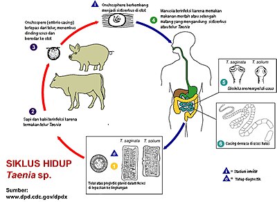
Telur taenia solium
| Taenia | ||||||||||||
|---|---|---|---|---|---|---|---|---|---|---|---|---|

Cacing Taenia saginata dewasa
| ||||||||||||
| Klasifikasi ilmiah | ||||||||||||
| ||||||||||||
| Spesies | ||||||||||||
Taenia crassiceps
Taenia pisiformis Taenia saginata Taenia solium Taenia asiatica Taenia taeniaeformis |
Taenia merupakan salah satu marga cacing pita yang termasuk dalam Kerajaan Animalia, Filum Platyhelminthes, Kelas Cestoda, BangsaCyclophyllidea, Suku Taeniidae. [1] Anggota-anggotanya dikenal sebagai parasit vertebrata penting yang menginfeksi manusia, babi, sapi, dan kerbau. [1]

Terdapat tiga spesies penting cacing pita Taenia, yaitu Taenia solium, Taenia saginata, dan Taenia asiatica. [2][3] Ketiga spesies Taenia ini dianggap penting karena dapat menyebabkan penyakit pada manusia, yang dikenal dengan istilah taeniasis dan sistiserkosis.[2]. Adapun perbedaan antarspesiescacing pita Taenia dapat dilihat pada Tabel 1 di bawah ini.
Tabel 1. Perbedaan antara Taenia solium, Taenia saginata dan Taenia asiatica
| No. | Keterangan | Taenia solium [1][4] | Taenia saginata [1][4] | Taenia asiatica [5] |
|---|---|---|---|---|
| 1 | Inang definitif danhabitat | Usus halus manusia | Usus halus manusia | Usus halus manusia |
| 2 | Inang antara | Babi dan manusia | Sapi (utama), kambing,domba | Babi (utama), sapi |
| 3 | Nama tahap larva | Cysticercus cellulosae | Cysticercus bovis | Cysticercus t.s. taiwanensis |
| 4 | Ukuran panjang xlebar | (3-8)x 0,01 meter | (4-15) x 0,01 meter | 4-8 meter |
| 5 | Jumlah segmen | 700-1000 | 1000-2000 | 712 |
| 6 | Jumlah telur | 30.000-50.000 di setiap segmen | lebih dari 100.000 di setiap segmen |
Endolimax

| Endolimax | |
|---|---|
| Scientific classification | |
| Domain: | Eukaryote |
| Kingdom: | Amoebozoa |
| Phylum: | Archamoebae |
| Genus: | Endolimax |
Endolimax is a genus of amoebozoa[1] that are found in the intestines of various animals, including the species E. nana found in humans. Originally thought to be non-pathogenic, studies suggest it can cause intermittent or chronic diarrhea.[2][3] Additionally, it is very significant in medicine because it can provide false positives for other tests, notably the similar species Entamoeba histolytica, the pathogen responsible for amoebic dysentery, and because its presence indicates the host has consumed fecal material. It forms cysts with four nuclei which excyst in the body and become trophozoites. Endolimax nana nuclei have a large endosome somewhat off-center and small amounts of visible chromatin or none at all.
| Entamoeba histolytica | |
|---|---|
 | |
| Entamoeba histolytica cyst | |
| Scientific classification | |
| Domain: | Eukaryota |
| Phylum: | Amoebozoa |
| Class: | Archamoebae |
| Order: | Amoebida |
| Genus: | Entamoeba |
| Species: | E. histolytica |
| Binomial name | |
| Entamoeba histolytica Schaudinn, 1903 | |
Entamoeba histolytica is an anaerobic parasitic protozoan, part of the genus Entamoeba.[1] Predominantly infecting humans and other primates, E. histolytica is estimated to infect about 50 million people worldwide. Previously, it was thought that 10% of the world population was infected, but these figures predate the recognition that at least 90% of these infections were due to a second species,E. dispar.[2] Mammals such as dogs and cats can become infected transiently, but are not thought to contribute significantly to transmission.
| Escherichia coli | |
|---|---|
 | |
| Scientific classification | |
| Domain: | Bacteria |
| Kingdom: | Eubacteria |
| Phylum: | Proteobacteria |
| Class: | Gammaproteobacteria |
| Order: | Enterobacteriales |
| Family: | Enterobacteriaceae |
| Genus: | Escherichia |
| Species: | E. coli |
| Binomial name | |
| Escherichia coli (Migula 1895) Castellani and Chalmers 1919 | |
| Synonyms | |
Bacillus coli communis Escherich 1885
| |
Tiada ulasan:
Catat Ulasan